Orthopaedic Surgery Research
The Department of Orthopaedic Surgery includes a robust research program that brings together an internationally recognized network of scientists—from across the University and at partner institutions—to elucidate mechanisms of pathogenesis of musculoskeletal disease, create innovative technology for cutting-edge musculoskeletal research, and develop novel strategies to improve the diagnosis, treatment and prevention of musculoskeletal conditions. While discovery is at the heart of what the scientists do, they are also focused on translating their findings into clinical solutions for patients in need of orthopaedic care. Those efforts are bolstered by the involvement of practicing surgeons who aim to develop new treatment strategies and diagnostic tools that can improve pain and function, leading to better outcomes for patients with musculoskeletal conditions and diseases.
Research Faculty
Research Cores and Labs
Bone Imaging Core Shared Resource
Musculoskeletal imaging
Summary: State-of-the-art musculoskeletal imaging and analytical technologies to generate, analyze, and interpret bone-imaging data. Also provides training in bone imaging and supports educational activities.
Contact Email: JDelgadocalle@uams.edu
Key Resources: Faxitron UltraFocus with Dexa, 2 Scanco microCT 40, Zeiss Axioimager M2 with motorized stage
Director: Jesús Delgado-Calle, Ph.D.
Location: Multiple
Further Detail: https://medicine.uams.edu/cmdr/cores/technical-cores/
The Bone Imaging Core, directed by Dr. Jesus Delgado-Calle, is located in the Winthrop P. Rockefeller Cancer Institute, 4th floor, and provides essential and varied bone imaging services to UAMS researchers, including X-ray, dual-energy X-ray absorptiometry, micro-Computed Tomography, bioluminescence and fluorescence imaging, and histomorphometry analysis. The facility offers consultation, training, technical advice, and collaboration, thus promoting cutting-edge science and serving as a central common area for investigators to interact and exchange scientific information. Bone imaging activities are carried out by a full-time employee, Stuart Berryhill, or investigators who are well-qualified operators and have received extensive training.
The UAMS Center for Osteoporosis and Metabolic Bone Diseases (CMDR), is a 15,000 square-foot facility in the Winthrop P. Rockefeller Cancer Institute funded by an NIH Centers of Biomedical Research Excellence (COBRE) grant. The CMDR is dedicated to the study of bone diseases and its treatment, houses one of the largest research units of its kind in the United States and is nationally and internationally recognized as a center of excellence in bone biology. The UAMS Skeletal Biology Group is one of the most diverse and accomplished in the country. Career levels span from the very young to the highly accomplished. Areas of expertise include aging, cancer in bone and metastasis, mechanobiology, endocrine function, fracture healing, and many other areas. The UAMS bone group has several senior faculty, including Drs. Weinstein, O’Brien, Almeida, Ambrogini, Morello, Yacobby, and Zangari, and has recently added to the group more senior faculty, Drs. Zhan, Bellido, Dr. Delgado-Calle, and several junior faculty, including Drs. Xiong, Kim, Onal, Dole, Sato, Noval, and Porter. In conjunction with the strong pre-existing bone faculty at UAMS, these new additions have resulted in a world-class bone group that exhibits robust depth and breadth of expertise in skeletal science, complemented by a rich track record of mentoring and leadership. Investigators employ a wide variety of cutting-edge technologies, and there is a culture that encourages highly interactive collaborative efforts. There are multiple interactions among the bone investigators on a weekly or daily basis through investigator meetings, journal clubs, and seminars. The CMDR houses four cores, including a Genetic Models Core that uses cutting-edge approaches to manipulate cells and mice genetically, a Bone Histology, Biomechanics, and Human Tissue Core that collects and processes histological samples from mouse and human skeletal tissues, a Bone Imaging Core that performs imaging and digital analysis of skeletal tissues, and a Bone Bioinformatics Core that analyzes the large datasets generated by the molecular analyses of bone cells from mouse and human samples.
The Bone Imaging Core employs a full-time technician. The Core houses two Dexa InAlyzer2, model M, scanners (MicroPhotonics) to analyze bone mineral density and body composition, and one UltraFocus Faxitron (Hologics) for digital x-ray imaging of live animals and excised tissues. For micro-computed tomography (microCT), the Core houses two vivaCT80 for live imaging and two CT45 for ex vivo imaging (Scanco Medical). Features include but are not limited to: a) scout view with calibrated diameter positions, b) single-slice and multi-slice 3D scanning (interactive slice number selection), and c) single or multiple sample scanning. Fast image reconstruction is available both online and offline. Analysis options include image analysis software, bone morphometry software, 3D morphometric analysis of trabecular and cortical bone, and 3D image rendering and visualization. The instruments are interfaced with an HP Integrity Server rx2660 (Dual Core-2 x 1.4 GHz CPU), 2 x 8 GB RAM (Max. 32 GB), 2×146 GB + 4×300 GB Disks, 2 x 10/100/1000 Mb/s Ethernet, and an HP 24” monitor. The Core also houses one IVIS Lumina XR Inst, Series III for bioluminescence and fluorescence imaging of live and excised tissues, as well as tissue cultures. The Bone Imaging Core has two separate microscope stations (bright field, epifluorescence, reflected bright field and dark field, and reflected and transmitted DIC) and two separate software packages for performing morphometric analyses (ImagePro Plus and Osteometrics image analysis system). Attached to the microscopes is a digitally cooled CCD micropublisher camera for taking high-resolution images. An 8-slide motorized stage from ASI enables automated image scanning to save time in image acquisition. The microscope and the automated stage are linked with Osteometrics bone analysis software (version 13.2).
Histology, Biomechanics, and Human Tissue Core Shared Resource
Hard tissue histology, and in vivo and ex vivo biomechanical testing
Summary: Highly specialized instrumentation and expertise for biomechanics and analysis of murine bones, human bone biopsies, and human joints.
Contact Email: JXIONG@uams.edu, EAmbrogini@uams.edu
Key Resources: Electroforce 5500, MTS machines, microtomes, embedding, vacuum ovens, multiple capabilities in histology staining
Director: Jinhu Xiong, Ph.D., Elena Ambrogini, M.D., Ph.D.
Location: Multiple
Further Detail: https://medicine.uams.edu/cmdr/cores/technical-cores/
The Histology, Biomechanics, and Human Tissue (HBH) Core of the Center for Musculoskeletal Disease Research (CMDR) provides state-of-the-art histology and biomechanical testing services for skeletal phenotyping. Located in ~2,000 sq. ft. of dedicated space on the 9th floor of the WRCI, the Core offers facilities for frozen sectioning, paraffin and methyl-methacrylate embedding, histological staining, and biomechanical testing. Key instrumentation includes three TA ElectroForce® 5500 test systems for mechanical property analysis of mouse bone, enabling three-point bending, ex vivo strain measurement, and in vivo tibial dynamic loading. Tail-suspension cages are also available to model disuse-induced bone loss in mice. Additional resources include paraffin-embedding stations, vacuum ovens, cryotomes, microtomes, optical microscopes, two Leica CM3050 S cryostats with CryoJane Tape Transfer System, and a diamond wire saw for plastic cross-sectioning. The Core also provides access to human bone samples for histology, in vitro cell culture, and gene expression studies, creating a translational bridge between animal models and human disease. By integrating advanced histopathology, functional biomechanics, and annotated human biospecimens, the HBH Core supports investigators in elucidating mechanisms of skeletal disorders and identifying novel therapeutic targets.
Epredia/Fisher HM355S Microtome (x4)
The Core houses four HM355S Microtomes to support diverse histological applications. Two instruments are dedicated to longitudinal sectioning of plastic-embedded specimens, enabling precise preparation of thin hard tissue sections (5 µm). The third microtome is reserved exclusively for generating RNase-free paraffin sections, ensuring high-quality RNA preservation for downstream molecular analyses. The fourth microtome is for regular paraffin sectioning of soft tissues and decalcified bone samples.
Leica CM3050S
The Core is equipped with Leica CM3050S Cryostats integrated with the CryoJane® Tape-Transfer System, enabling the preparation of high-quality frozen tissue sections with superior structural preservation. The tape-transfer technology minimizes artifacts and distortion, allowing reliable sectioning of delicate or challenging specimens such as bone.
Sakura GSI-128 Paraffin embedding station
The Sakura GSI-128 Paraffin Embedding Station provides a fully integrated platform for the preparation of paraffin-embedded tissue specimens. The system combines a heated paraffin reservoir, embedding module, and cold plate to ensure consistent sample orientation and rapid solidification. Programmable temperature controls maintain optimal conditions for both paraffin and molds, minimizing variability and preserving tissue integrity. The GSI-128 enables high-throughput embedding of tissue blocks, supporting downstream histological sectioning and staining.
Applied Diamond, INC. 3241 Precision Diamond wire saw
The Applied Diamond 3241 Precision Diamond Wire Saw is designed for thick cross-sectioning of plastic-embedded specimens. This system enables the preparation of high-quality sections in the range of 70–200 µm, suitable for quantification of periosteal bone formation rate.
Buehler IsoMet Low Speed Saw Cross sectioning saw (x2)
The Applied Diamond 3241 Precision Diamond Wire Saw is designed for thick cross-sectioning of plastic-embedded specimens. This system enables the preparation of sections in the range of 100–200 µm, suitable for quantification of periosteal bone formation rate.
Patterson Brand Grinding Machine
The Core utilizes a Patterson Brand Grinding Machine to prepare plastic-embedded tissue blocks for sectioning. This instrument is used to trim and grind blocks to the proper size and shape, ensuring secure mounting and optimal alignment for subsequent microtome sectioning. By producing smooth, uniform block surfaces, the grinding machine facilitates precise longitudinal or transverse sectioning of hard tissues and resin-embedded specimens.
Buehler 49-10070 Grinding Machine
The Core is equipped with two Buehler 49-10070 Grinding Machine to polish thick cross-sections. This system produces smooth, flat surfaces on plastic-embedded specimens, ensuring high-quality sample preparation for light and polarized microscopy. By removing surface irregularities and achieving uniform finishes, the grinding machine facilitates accurate visualization and structural analysis of hard tissues.
Waters TA Electroforce 5500 Mechanical testing machine
The Core houses three Waters TA ElectroForce® 5500 mechanical testing systems to support functional assessment of mechanical properties of murine bone. One system is configured for three-point bending to evaluate mechanical strength and stiffness of bone specimens. The other two systems are dedicated to in vivo tibial dynamic loading, enabling controlled application of cyclic mechanical forces to the mouse tibia for studies of mechanotransduction and bone adaptation. Together, these instruments provide versatile, high-precision platforms for both ex vivo biomechanical testing and in vivo loading experiments.
General laboratory equipment
• Incubator
• Lighted Tissue Bath
• Tissue Floating bath
• Slides warmer
• Vacuum Oven
• Fisher Scientific FW02-07-4121 Microscope
• Olympus SZ-ST Microscope
• Olympus Microscope
• Fisherbrand Microscope
Dan Snider and Jon Zieske UAMS Golf Lab
The Dan Snider and Jon Zieske UAMS Golf Lab is located inside the 5,000-square-foot UAMS Biomechanics Lab at 1701 Aldersgate Road, where UAMS researchers conduct clinical and biomechanical research to help physicians better understand how patients move before and after orthopaedic surgical procedures and treatments. C. Lowry Barnes, M.D., UAMS Chancellor, opened the facility as his private laboratory in 2010. When he came to UAMS in 2014, it became the UAMS Biomechanics Lab. Researchers have used the lab in a wide variety of gait, balance and movement studies, including those involving yoga and ballet, with golf-focused research added later.
The golf lab features advanced technology and state-of-the-art equipment to analyze and improve golf swings, prevent injuries, and support rehabilitation.
3D Motion Analysis:
The motion capture system, complete with infrared cameras, records subjects as they perform various movements. It measures exact joint angles, swing mechanics, and body movements to maximize efficiency.
Force Plate Technology:
Force plates embedded in the floor evaluate weight shifting, measure ground reaction forces and balance during a swing to create a comprehensive, real-time picture of the mechanics of swinging a golf club.
Interactive Golf Simulator:
Participants hit golf balls from an artificial turf, and the system calculates ball speed, launch angle and the distance the ball would travel on a real golf course. Subjects can play many golf courses around the world.
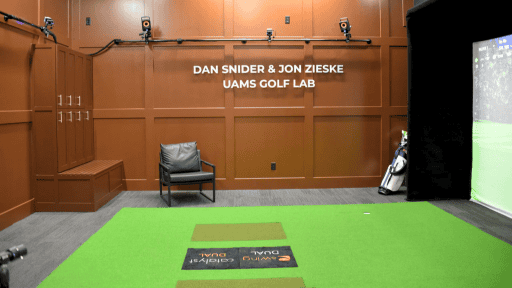
Inside the golf lab is an interactive golf simulator that calculates ball speed, launch angle and the distance the ball would travel on a real golf course.
Center for Musculoskeletal Disease Research (CMDR)
Razorback Research Catalyst

Hip and Knee Surgery Research All-Stars
We are proud to be named among the Avant-garde Health Hip and Knee Surgery Research All-Stars for 2025. UAMS Hip and Knee Joint Replacement Surgeons C. Lowry Barnes, M.D., Jeffrey Stambough, M.D., and Benjamin Stronach, M.D., and Simon Mears, M.D., Ph.D. are among the top 3% of Hip and Knee Surgeons named as Research All-Stars for 2025 based on publication volume, journal impact factor, and author position.
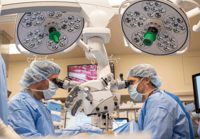
Delivering Innovation for Impact
UAMS orthopaedic hand surgeons John Bracey, M.D. and Mark Tait, M.D. along with neurosurgeon Erica Petersen, M.D. implant innovative prosthetic hand, developed by researchers at the Institute for Integrative and Innovative Research (I³R), that restores a meaningful sense of touch and grip force following surgery at the University of Arkansas for Medical Sciences (UAMS). Read more